
2017
We have published a new data analysis method, called skCSD, to reveal membrane currents on single neurons, based on extracellular multichannel electrode array measurements. The new method provides higher precision in membrane current source density reconstruction due to the inclusion of the morphology information into the calculation. We have applied the new method to the first available parallel extracellular and intrancellular data and showed the spatial propagation of the currents during action potential generation on the dendritic branches of the cell. (Fig. 1) The scripts for the analysis, written in R, were tested and made publicly available as an open source program package.
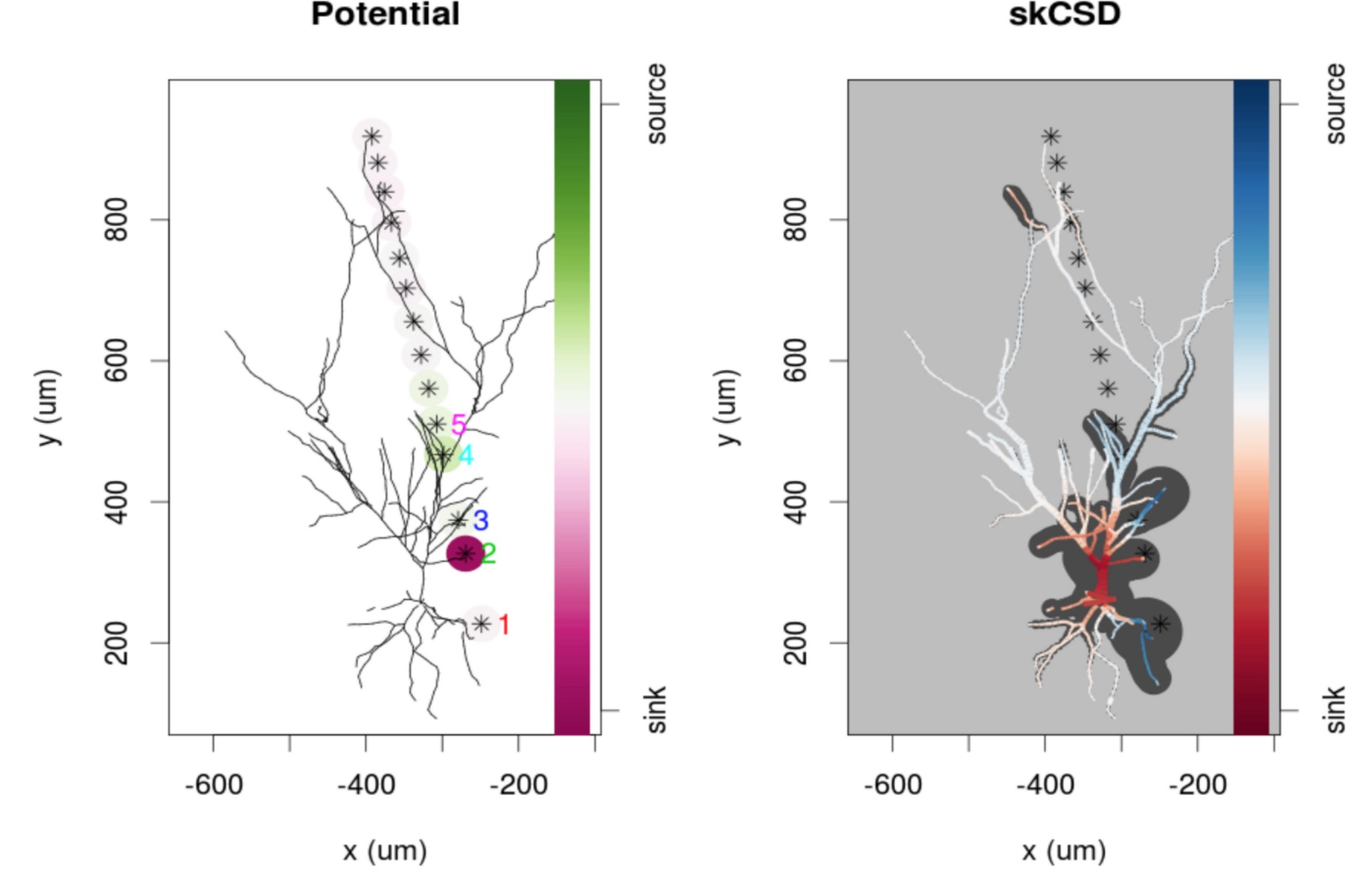
Figure 1: Left: Electrode positions (stras) and reconstructed morphology of the pyramid cell from the CA1 region of the rat hippocampus. The color coded circles on the electrodes show the measured momentary electric potential at the moment of the peak of an action potential generated by the neuron. Right: The reconstructed current source density distribution along the dendritic tree of the neuron. Warm colors mark inward positive currents to the neuron (sink) cold colors mark the outward currents from the neuron (sources).
We have published the first results on the analysis of the parallel recordings on intrinsic optical and local field potential by a transparent electrode array. The new method makes possible the fusion of the two methods, by exploiting the advantages of both, the excellent spatial resolution of the optical imaging and the excellent temporal resolution of the electric signal.
We applied our coherence clustering method, to determine the cortical structures and areas from the measurements with the transparent cortical surface electrode grid, parallel to the intrinsic optical signal measurement. The methodology and the first results were published on a conference and in a proceedings journal.
We created a new feedback model of the dynamics of gene expression and protein synthesis on the basis of experimental findings. We built a stochastic kinetic model to investigate and compare the “traditional” and the feed-back model of genetic expression processes. Qualitative and quantitative changes in the shape and in the numerical characteristics of the stationary distributions of proteins and RNA molecules suggest that more combined experimental and theoretical studies should be done to uncover the details of the kinetic mechanisms of gene expressions.
We showed that in the somatosensory cortical circuitry, which is largely responsible for tactile perception, lateral interactions mostly depend on the intra-areal connections complemented by the neuronal feedback originating from areas with higher order functional representations. In contrast, feedforward connections from lower order areas exhibit spatially restricted lateral spread indicating higher functional specificity. Our results also suggest that the population activity is mostly determined by the target regions of the feedforward connections overlapping the strong local input within an area. The manuscript including these findings has been submitted for publication and is now under major revision.
To better understand somatosensory, and in general cortical communication, we studied the synaptic organization of the above mentioned connections in 3D by way of electron microscopy. Using state of the art data analyses techniques we found that the size of axon terminals is an important distinguishing morphological feature of the cortical synapses. We also found that the size of the mitochondria and postsynaptic densities (the active zone of the signal transmission) relative to the size of the axon terminals also exhibit important distinguishing characteristics and that their positive correlation can be explained by the energy need of synaptic transmission. The manuscript summarizing these findings is going to be submitted soon.
Our ongoing studies show that the robustness and synchronizability of the network of cortical areas is especially sensitive to targeted removal of the network edges on the basis of their convergence degree introduced previously by our group.
By including a Bayesian evaluation algorithm, the development of our new causality analysis method, now we call it dimensional causality method (DC), has been completed. The DC method has been tested on various simulated systems, such as coupled Lorentz systems, coupled logistic maps and coupled Hindmarsh-Rose models. These simulated dynamical systems pose different challenges towards the DC algorithm but we found, that all the DC method was able to infer all possible causal relations (unidirectional, circular, independent and hidden common cause) in all the three model cases. Preliminary applications were made on neurophysiological data from epileptic patients, during photostimulation experiment and during epileptic seizure.